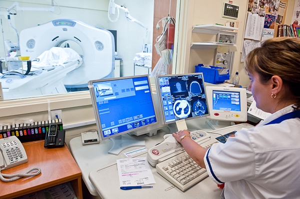
Radiology (X-ray) is a medical specialty which uses a variety of different imaging technologies and equipment to image the body and diagnose disease.
These technologies include X-ray, Magnetic Resonance Imaging (MRI), Computed Tomography (CT), Ultrasound, Nuclear Medicine and Positron Emission Tomography (PET).
Our radiology department is located on the first floor of the Clinical Services Block via the hospital’s main entrance. Directions are signposted.

| Contact Us | Opening Hours | ||||||
| Radiology reception | 06 348 1308 | X-ray GP patients | 8am – 4.30pm, Mon-Fri | ||||
| Ultrasound bookings | 06 348 3224 | X-ray OPD patients | 8am – 4.30pm, Mon-Fri | ||||
| CT/MRI bookings |
06 348 3303 |
X-ray ED/WAM | 8am – 9pm, Mon-Fri 10.30am - 6pm, Sat/Sun |
||||
|
Xray bookings |
06 348 3319 |
||||||
| CT | 8am – 5pm, Mon-Fri | ||||||
| Complaints | MRI | Mon & Fri, 8am – 4pm Tues, Wed & Thurs, 7am - 9pm |
|||||
| Radiology Clinical Manager | 06 348 3346 | Ultrasound Please note: muscular skeletal ultrasound (e.g. shoulders, elbows, knees) cannot be performed at Whanganui DHB. |
8am – 4pm |
||||
| Customer Relations Officer | 06 348 3045 | ||||||
| On-call emergency services | |||||||
| An emergency service for X-ray, Computerised Tomography (CT) and Ultrasound(US). | |||||||
Our department
Our radiology department is equipped with the latest imaging equipment including:
- one x-ray room in the main department and one in the emergency department
- Fluroscopy
- Computed Tomography (CT)
- two general Ultrasound rooms - Please note: muscular skeletal ultrasound (e.g. shoulders, elbows, knees) cannot be performed at Whanganui DHB.
- an echocardiography ultrasound room
- Magnetic Resonance Imaging (MRI)
- Image Intensifier (II) for use in theatre, two mobile x-ray machines for critically ill patients in the emergency department and the wards.
Please note
All CT, MRI and Ultrasound patients are sent out appointment times. The schedule is tight so please arrive at the time indicated on your appointment letter. If you arrive late your appointment may need to be re-booked. Emergency cases need to take precedence so please be aware you may be required to wait a little longer.
Should you be unable to attend your appointment, please contact us as soon as possible. If you are unable to attend your appointment, and do not contact us, you will be removed from the waiting list and your referral sent back to your doctor.
Off-site services (certain criteria apply):
- Mammography – Breastscreen Aotearoa, Palmerston North
- Bone Densitometry – Cortex Medical Imaging, Whanganui
All examinations/images are reviewed by a radiologist. A report is then written and sent to your consultant/doctor.
The report is usually available to your doctor within five working days. Should the radiologist find anything requiring urgent attention, your doctor will be contacted.
The team
Radiologists
Radiologists are specialist doctors who read and understand your films. They will also be involved if you have a barium study (a specialised x-ray exam that uses a special dye which is visible on x-rays and highlights your anatomy) or a CT/US guided biopsy. Radiologists interpret the results of the images and send them to your doctor.
Te Whatu Ora Whanganui has one full-time, on-site radiologist with Pacific Radiology (based in Wellington) also contracted to report on examinations.
Medical Radiation Technologists (MRTs) or Radiographers
MRTs perform your x-ray, CT or MRI examination and send the images through to the radiologist for diagnosis.
In New Zealand, MRTs are required to complete a three-year medical imaging degree before they can practice. Once qualified they are required to be registered in an independantly-assessed continued professional development (CPD) program every three years.
Student MRTs from UCOL also work in the department as part of their practical training. They will always identify themselves to a patient and ask if they are comfortable with them performing the examination. If you do not wish for a student to perform your exam please let us know and a qualified MRT will be available to help you.
Sonographers
Sonographers are MRTs specially trained to operate ultrasound equipment, interpret images and provide initial findings. They are required to have completed a post-graduate qualification in ultrasound.
Radiology nurses
Radiology nurses are registered nurses who help the radiologists and MRTs perform interventional procedures in CT, ultrasound and screening. They are also available should you suddenly feel unwell while visiting the department
Technologies/equipment
Te Whatu Ora Whanganui has one general x-ray room within the Radiology Department, as well as one in the hospital’s Emergency Department. A screening room and Orthopantomogram (OPG) machine (a special scanning dental x-ray unit) are also based within the department.
All GP and outpatient x-rays are performed in the main department. A booking system is in place for x-ray appointments.
If you have small children or unable to wait, please feel free to call before leaving home so we can let you know the approximate wait times.
Emergency patients, Whanganui Accident and Medical (WAM) patients and inpatients are all imaged in the x-ray room located near the Emergency Department. If you are a patient in WAM or the Emergency Department, your doctor will bring us your x-ray request form and we will call you for your examination soon as possible.
The screening room
Fluoroscopy, or x-ray screening, is a specialised x-ray unit which allows the doctor to see x-ray images on a television in the room as the x-rays are taken.
Examinations using fluoroscopy usually require a radiologist to supply you a special dye that will show up brightly on the x-rays - this dye is called a contrast agent. This contrast agent can be swallowed or injected to show different parts of your body.
OPG machine
Te Whatu Ora Whanganui dental patients are often sent to the Radiology Department for imaging of their entire mouth taken with this machine. The OPG machine is used for this.
Computed Tomography (CT)
CT uses x-rays to produce detailed three dimensional images of the head and body. This is especially useful for looking at soft tissue structures such as lungs, liver, colon. Images are created when x-rays travel through the body to detectors within the scanner. Computer software takes measurements from the detectors and creates an image.
Length of examinations
Scans usually take 15-30 minutes. You may be asked to arrive up to 45 minutes prior to your appointment time to allow time for important preparation for the scan. For some scans we will ask you to drink water/contrast media when you arrive.
Depending on the scan required, you may be asked to get a blood test or follow a special diet a few days before your examination. A blood test request form or instructions will be sent to you.
What happens?
During the scan you will lie on a narrow table that moves through a circular opening. You may be asked to hold your breath at certain times but it is important to remain still. Slight claustrophobia is experienced by some patients. This is not common.
You may require an injection of contrast media during your scan, which is used to create clearer images and help with the diagnosis. A staff member will discuss this with you when you arrive. You will need to sign a questionnaire/consent form prior to the scan. Please bring your reading glasses.
Magnetic Resonance Imaging (MRI)
MRI uses magnetism and radio frequency to produce detailed cross-sectional images of the body part being examined. MRI is used to show soft tissue structures within the body, although it can also be used to image bone.
MRI uses a strong magnetic field to help produce the images. Because the high magnetic field strength is potentially dangerous therefore everyone entering the scan room needs to be carefully screened to ensure their safety. You will be sent a questionnaire with your appointment letter. Please complete the questionnaire before attending your examination, and bring your reading glasses with you.
Please contact us for further checks to ensure you can be scanned safely, if you have any of the following:
- a cardiac pacemaker.
- are in the first three months of pregnancy.
- have certain aneurysm clips, heart valves
- have metal fragments embedded in the eye or body.
Length of examinations
MRI scans usually take between 30 and 60 minutes, however more complex examinations can take up to 90 minutes. Some examinations may require that you do not eat before your examination or that you follow a special diet. Your appointment letter will include all of this information.
Depending on the scan you are having, you may be asked to get a blood test a few days before your examination. A blood test request form will be sent to you.
What happens?
You will be required to lie flat on a narrow table inside the MRI machine. A MRI coil will be placed around the area to be imaged. In order for us to obtain high quality images, it is important that you are completely still during the examination. Movement can blur the images.
Because the machine is noisy, you will be given some ear plugs or headphones to wear and a panic button to hold just in case you need to get the MRT’s attention. The MRT will be able to talk to you though an intercom during your exam.
If you bring a CD with you, we can play it through the headphones. (Please note that iPods and MP3s will not work).
An injection of contrast media may be required during your scan. This is used to create clearer images and helps with diagnosis. A staff member will discuss this with you when you arrive.
If you are claustrophobic, please call us as soon as you receive your appointment. It may be necessary for you to get some sedation medication from your GP. You are welcome to bring a support person with you, but please note they will also need to complete the MRI safety questionnaire.
Ultrasound - Please note: muscular skeletal ultrasound (e.g. shoulders, elbows, knees) cannot be performed at Te Whatu Ora Whanganui.
Ultrasound images are produced by using high frequency sound waves too high for us to hear. Ultrasound is produced within a hand-held transducer/probe placed on your skin. As the sound waves are produced, they pass though your body and then return to the transducer. The computer converts these to images which are displayed on the monitors.
Preparation
Your preparation will depend on the nature of your examination. Details/instructions will be sent to you with your appointment. It is really important that you follow the instructions, otherwise we may not be able to perform the examination.
Length of examination
The examination usually takes between five and 45 minutes. This will depend on the type of ultrasound you have been referred for.
What happens?
You will be asked to lie down on the bed/couch to allow us to access the area to be scanned. Gel will be placed on your skin with the ultrasound probe placed on top. It is sometimes necessary to add pressure to improve the image quality.
Support person
Patients are welcome to bring a support person with them.
Frequently Asked Questions
Why do I need to confirm my appointment?
Due to the high demand of our service, it is important that the machines are used efficiently, and for the maximum amount of time during the day.
If you do not confirm your appointment extra time is spent trying to contact you. If you are not going to attend we can give your appointment time to someone else.
When will I get my results?
Once your examination is completed, the images are sent to a radiologist to review and write a report. This report will be sent to your doctor.
Your doctor (GP/specialist) will, usually, receive the results about five days after the examination. Your doctor will let you know the results of your examination. You should contact your doctor if you have not heard from them within ten days of your examination.
Why can the MRT not tell me the results of my x-ray/scan?
MRT’s are trained to take the images and the radiologist has the specialist skills required to interpret the images. It would be inappropriate for a MRT to offer an opinion about your image as they do not have the skills or training to read the images.
I am pregnant. Is it safe for me to have a x-ray/scan?
This depends on what and why you need the examination.
For x-ray and CT, we always try to limit your exposure to radiation. This is especially important when a patient is pregnant. However, the risk of not accurately diagnosing and treating your condition is sometimes more harmful than the risk of the radiation.
With MRI patients, we prefer not to scan those who are pregnant. However, if a scan cannot be postponed until after birth, the patient must be 14 weeks pregnant or more.
Women can safely have an ultrasound at any stage during pregnancy.
Can I eat and drink normally prior to my scan?
This depends on the type of examination you require. If you are to stop eating or follow a special diet, you will receive the instructions with your appointment letter.
Should I stop taking my medication before my exam?
No, unless you are having a MCRP (gall bladder study) in which case, bring them with you to take immediately after the examination.
Can I bring a support person with me?
X-ray and CT: Because of the radiation exposure during these examinations we do not allow support people in the room unless absolutely necessary. If you need a support person to stay with you in the room during the examination, we will give them a lead coat to wear.
MRI: For safety reasons we limit the number of people in the scan room to those essential for the examination. However you can have one support person with you during the scan if needed. A support person must accompany children under 12. The support person will need to complete a safety questionnaire as well.
Ultrasound: While there are no safety reasons for limiting the number of people allowed in the room when you have your scan, we do ask that you limit the number of people you bring with you. It is important that the sonographer is able to concentrate while performing your examination.
Please note
Some examinations can take 45-60 minutes. If you need to bring small children with you, please bring some entertainment for them as our staff will not be able to look after your children while you have your examination.
I am pregnant. Can I be a support person?
Yes, but only for an ultrasound examinations.
Will my valuables be safe in the changing room?
Yes. All changing rooms, apart from in the CT room, can be locked or are situated in restricted areas. CT patients will be asked to bring their valuables in to the scan room with them
Should I leave my hearing aids at home?
If you need hearing aids to hear general conversation adequately, please bring them with you. The MRT will advise you if they need to be taken out.
Should I bring my reading glasses?
Yes. You may be required to read some information and complete a consent form.


